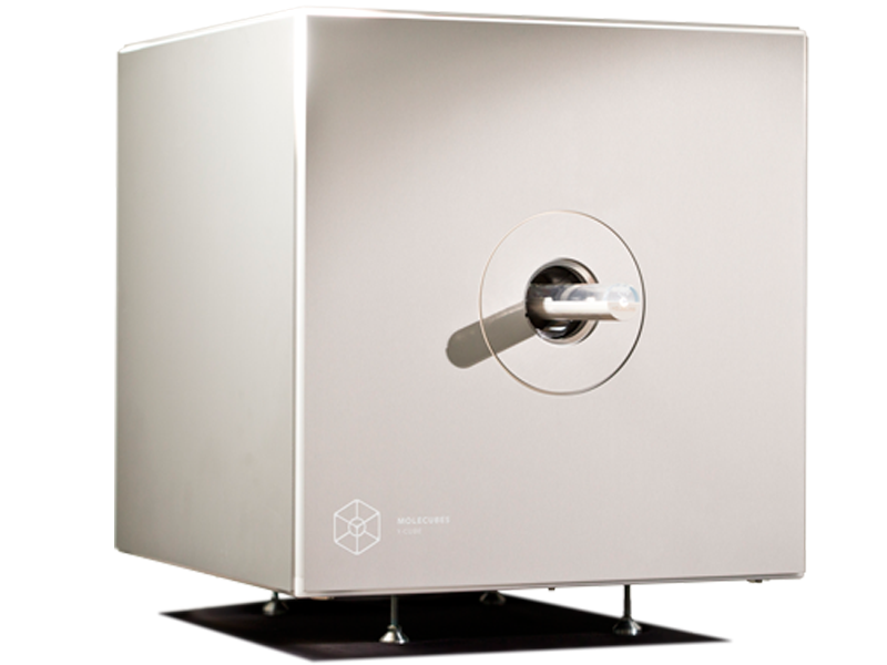
The X-CUBE is a high throughput preclinical CT imager enabling in-vivo tissue quantification

The X-CUBE allows for fast whole body mouse and rat CT imaging at extremely low dose and excellent soft tissue contrast. Light weighted thanks to a self-shielded imaging unit it is a truly mobile in vivo scanner.

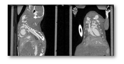
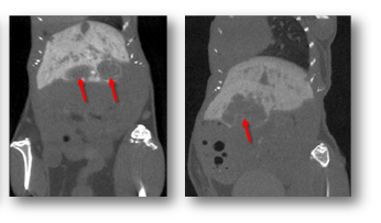
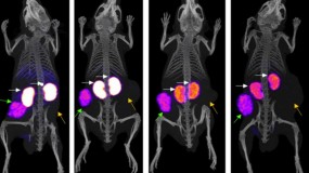
CT imaging enables a wide range of non-invasive and longitudinal tissue measurement applications in your preclinical models. These include orthotopic and subcutaneous tumour volume, tumour metastases volume, lung volume, heart ejection fractures and blood pool volume, and more.

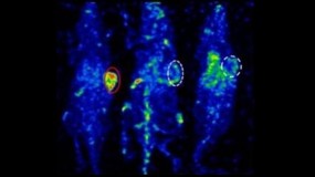
Intuitive and wireless software combined with our multimodal small animal bed allow for easy and modular multimodal imaging along with the γ -CUBE (SPECT) and β -CUBE (PET).

X-CUBE preclinical CT imager testimonials


γ-CUBE

X-CUBE

β-CUBE

  GP mouse protocol    
Field Of View
axial x transaxial
12mm x 30mm 35mm x 63mm 130mm x 72mm
Spatial Resolution
*general purpose mouse collimator
< 0,6mm 0,05mm < 1mm
Sensitivity 0,12% - > 10%
Reconstruction Code
on board GPU-based
3D MLEM, 3D OSEM FDK, ISRA FBP, 3D MLEM, 3D OSEM
Weight
over footprint of 54cm x 54cm
< 80kg < 100kg < 90kg

X-CUBE existing customers 

Click on the tabs below to find out more about our customers, and how they are using the X-CUBE in their laboratories.

Kyoto Pharmaceutical University, Japan

Studying the biodistribution of Eph receptors for cancer diagnosis

A study published by Furukawa et al. at Kyoto Pharmaceutical University has used MOLECUBES’ X-CUBE and γ-CUBE to study the biodistribution of Eph receptors, which are thought to be associated with cancer cell migration and proliferation. The benchtop preclinical imagers were used to evaluate and visualise a EphA2 receptor-specific SPECT imaging tracer for diagnosing cancer called [I-125]ETB.

You can learn more about the study and how the MOLECUBES were used by clicking the button below.

READ MORE

BIOEMTECH, Greece

BIOEMTECH, Greece

Prof. George Loudos is the founder and CEO of BIOEMTECH. BIOEMTECH labs, are located in Athens and support the complete chain of pre-clinical studies for testing new  compounds  including: animal hosting premises (mice and rats), in vitro lab for cell studies, radiochemistry lab for labelling various compounds and multimodal imaging facility,  including fast  dynamic molecular screening, 3D SPECT/CT systems (x/γ-CUBE, Molecubes), plus a PET and optical system, developed in house. 

Why choose Molecubes?

BIOEMTECH has an academic origin and deep technical background, since we have launched the first in vivo molecular screening systems (the eyes) for whole body planar dynamic imaging. We chose Molecubes because of the teams’ academic background and thus deep knowledge of the technology and applications. Knowing the core team and their progress for more than a decade, we felt from the first moment absolute trust in their proposed solutions but also in their support and innovative ideas.

How would you rate your experience with Molecubes?

Having two of the Molecubes systems for almost 2 years now, we have deeply appreciated the end user experience and the continuous support.

What specific feature do you like the most?

Some of the features we have mostly appreciated is the wide range of applications that can be covered, along with a great simplicity of use, beautiful images and accessibility and promptness of the supporting team.

Which applications do you run on the system?

Up to now, we have used the systems for multiple SPECT applications on cardiac imaging, several oncology applications, lung imaging, brain targeting and a wide range of nanoparticle studies. On CT, besides animals, we have imaged multiple stained tissues with great accuracy, as well as dental tissues and in vivo bone studies.

Would you choose for benchtop instruments again?

It is in our plans to also purchase the β-CUBE and thus have the complete CUBES platform in our pre-clinical labs.

How often are the instruments used?

The instruments are used on a weekly basis and during busy periods daily. Acquisitions and reconstructions are running in parallel for a higher throughput. Since our throughput is high, we look forward to seeing new  improvements  related to multiple animal imaging or remote workstations for parallel post-processing of the acquired data, greatly saving experimental time.

Infinity Lab, Ghent University, Belgium

Infinity Lab, Ghent University, Belgium

Prof. Christian Vanhove is responsible for the day-to-day functioning of the preclinical INFINITY lab and the coordination of the partner research, either from within Ghent University, from private pharma or biotech companies.

Why choose Molecubes?

The lab will move to a new building in a few years from now. Luckily, the benchtop modular system allows me to move all 3 modalities at no extra cost or time loss for recalibration. The new lab will have less space, so I’m very happy that these benchtop systems exist today
without compromise on image quality.

How would you rate your experience with Molecubes?

The PET, SPECT and CT hardware and software proved to be very robust and reliable over the past year. To date, we have already imaged over 100 mice and rats effortlessly. Whenever we have a new request from a partnering lab, we can reach out to Molecubes for questions related to the specific projects.

What specific feature do you like the most?

All 3 modalities provide 1 convenient DICOM file to work with, there is no manual interaction needed for co-registration and still all CUBES can be used in stand-alone mode when needed. The whole process is automated and produces publication-ready images. To me, that’s true flexibility!

University of Pennsylvania, USA

University of Pennsylvania (UPENN), USA

The Small Animal Imaging Facility (SAIF) provides in vivo functional and anatomical  imaging and image analysis for cells, tissues, and small animals, primarily mice and rats, but also primates.

The assets of the SAIF include state-of-the-art instrumentation and a nationally recognized staff. The lab has a MOLECUBES PET/CT imager installed since June 2017.

Why choose MOLECUBES?

“The high performance made MOLECUBES an obvious choice for us, together with the clever design and excellent engineering. In addition, the option to use the CUBES individually or together was an important factor since our goal was to increase throughput of our small animal imaging facility. The long standing academic relationship with Ghent Group and trust in their capabilities made us take the final decision.”

How would you rate your experience with MOLECUBES?

“Delivery and install were on time as promised and very professional. Training was short and very effective, and users are very pleased with the system. Considering that we have the first CUBES to be installed outside of Ghent it is impressive how reliable they have proven and how effectively MOLECUBES is able to remotely handle a few initial problems.”

What specific feature do you like the most?

“We appreciate the simple and intuitive user menu and operation of each CUBE both separately and together. Using micro CT for attenuation correction and registration with PET is straight-forward. Separate CUBES allows multiple researchers to work simultaneously. Overall the CUBES operate reliably allowing on time scheduling of studies from different UPenn research groups. Finally, MOLECUBES is open and eager to gather users’ input to improve on their current and future features.” 

NYU Grossman School of Medicine, USA

Imaging in a BSL-3 lab for infectious diseases

The Preclinical Imaging Laboratory at the NYU Grossman School of Medicine, as part of the Division for Advanced Research Technologies within the Office of Science & Research, enables NYU biomedical scientists to image live animals on an organ, tissue, cell, or molecular level using state-of-the-art preclinical imaging technologies. The lab is equipped with MOLECUBES benchtop preclinical imagers including a γ-CUBE for SPECT, a β-CUBE for PET, and two X-CUBEs for CT, one of which was originally procured specifically for infectious diseases research including COVID-19.

We spoke to Director of the Preclinical Imaging Laboratory and Associate Professor Youssef Wadghiri about his work using the CUBES. You can find out what he had to say by clicking the button below. 

READ MORE

Hull PET Research Centre, UK

Steve Archibald: Why Hull chose MOLECUBES’ preclinical imagers for translational studies

MOLECUBES preclinical imagers have been installed at the PET Research Center (PETRC) at the University of Hull in the UK. The state-of-the-art facility uses medical imaging for research and the early diagnosis of diseases such as cancer and dementia. It supports a wide range of internationally-recognised research programmes and is part of a group of integrated clinical facilities with links to Castle Hill Hospital.

PETRC’s lab is equipped with a full suite of MOLECUBES including a γ-CUBE for SPECT, a β-CUBE for PET, and a X-CUBE for CT. By combining all three CUBES, the limitation of each modality is offset and provides a multimodal approach to preclinical imaging.

Watch our video with Professor of Molecular Imaging Steve Archibald or click the button below to find out more.

READ MORE

Documents

The LabLogic Group will always treat your details with the utmost care and will never sell them to third party companies.

Please choose one of the following:


Previous
Next