Studying the biodistribution of Eph receptors for cancer diagnosis with MOLECUBES
16th January 2024
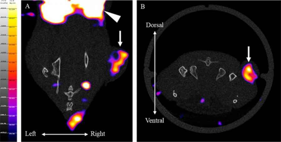
SPECT imaging of tumour-bearing mouse. Representative [I-123]ETB SPECT/CT imaging in tumour-bearing mouse at 205 min after injection: (A) dorsal view and (B) caudal view image. The tumour (arrow) and the kidney (arrowhead) are shown.
Kyoto Pharmaceutical University has been using the benchtop preclinical imagers to image a new SPECT radiotracer
A study published by Furukawa et al. at Kyoto Pharmaceutical University has used MOLECUBES’ X-CUBE and γ-CUBE to study the biodistribution of Eph receptors, which are thought to be associated with cancer cell migration and proliferation. The benchtop preclinical imagers were used to evaluate and visualise a EphA2 receptor-specific SPECT imaging tracer for diagnosing cancer.
Understanding the migration and proliferation of cancer cells
Erythropoietin-producing hepatocellular (Eph) receptors function as receptor tyrosine kinases involved in mediating cell-cell contact. Eph receptors, along with their ephrin ligands, are anchored to cell membranes, and their interaction leads to dimerization and subsequent activation. Based on these processes, it is hypothesised that the overexpression of Eph receptors in cancer cells plays a crucial role in their migration and proliferation when compared to normal cells.
Despite advancements in understanding Eph receptors, their detailed mechanisms are yet to be fully understood. Therefore, there is a need for a non-invasive method to measure the distribution and density of Eph receptors in tumours, providing valuable insights for cancer diagnosis and aiding in the development of therapeutic drugs.
Visualising implanted tumours using SPECT/CT imaging
A study conducted by Furukawa et al. aimed to address this gap by developing a small molecule SPECT imaging tracer called [I-125]ETB, specific to Eph receptors. The choice of SPECT as an imaging technique was justified by its high clinical versatility and widespread use in non-invasive imaging applications.
In this study, the γ-CUBE and X-CUBE were used as a non-invasive tool to evaluate and visualise the in vivo biodistribution of Eph through [I-125]ETB. SPECT/CT imaging was conducted on tumour-bearing mice in which [I-123]ETB was administered intravenously. A CT scan using a X-CUBE was initially performed for attenuation correction followed by a SPECT scan using a γ-CUBE.
As shown in the image above, the implanted tumours in the right limbs were clearly visualised in both dorsal and caudal images. Furthermore, in accordance with the ex vivo distribution data, a high accumulation of [I-123]ETB in the kidney was also observed.
Find out more
You can learn more about MOLECUBES by clicking the button below to request a guided demonstration with a product specialist or click here to read the full study.