PET/SPECT imaging in the development of targeted radiopharmaceuticals
29th June 2023
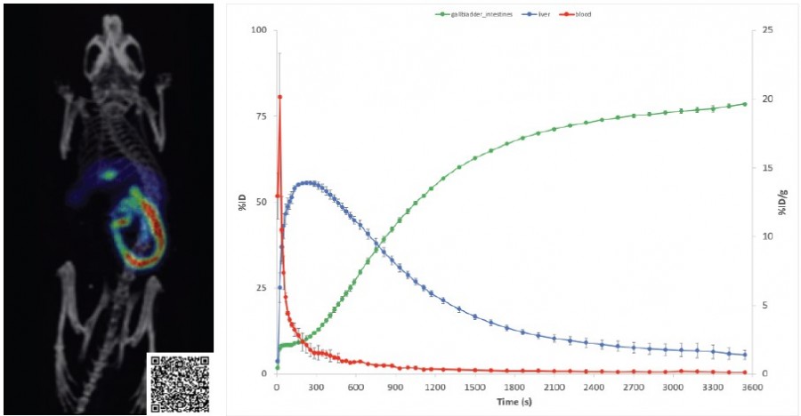
Dynamic radiotracer 18F-Fluorocholic Acid, a bile acid analogue providing quantitative kinetic information in multiple organs of interest.
MOLECUBES preclinical imagers allows modalities to be combined when studying the efficacy of new tracers
PET/SPECT imaging has proven advantages in the development of targeted radiopharmaceuticals. While anatomical imaging such as MRI and CT offer detailed structural information with high resolution, functional PET/SPECT imaging is highly sensitive and provides insights into the molecular biology of disease processes. Each modality has its own advantages and limitations, but these can be overcome by combining modalities, such as PET-CT or SPECT-CT. MOLECUBES benchtop preclinical imagers provide a multimodal approach with three models of CUBES available.
Using these compact benchtop imagers, MOLECUBES removes the barriers to preclinical PET, SPECT, and CT imaging whilst offering the highest performance standards supported by fast and simple workflows with a comprehensive understanding of the pathophysiological processes being studied.
In their white paper Advantages of small animal imaging in the development of targeted radiopharmaceuticals, MOLECUBES outline the advantages of PET/SPECT imaging to study disease/therapy progression when studying the efficacy of newly developed tracers.
The advantages of in vivo studies over ex vivo
When compared to cut and count (ex vivo) studies, PET/SPECT imaging allows full biodistribution data of all organs at multiple time points in the same animals. This reduces the number of animals necessary in a study, aligning with the principles of the 3Rs (Replacement, Reduction, Refinement) and in turn reduces the cost of study when using expensive animal models. It also provides all organ information in one dataset, meaning organs of interest do not have to be selected in advance. This is advantageous when developing new radiotracers, as it can be hard to predict the target organs using cut and count.
Furthermore, each animal serves as its own control in imaging studies, leading to a reduction in the standard error of the data. PET/SPECT imaging is a non-invasive in vivo technique, allowing researchers to evaluate animals at multiple time points before reaching the study endpoint. This feature enables earlier and time-dependent detection of therapeutic effects, enhancing the researcher's ability to assess the efficacy of treatments.
Find out more
You can learn more about the CUBES by clicking the button below to speak to a product specialist or by downloading the MOLECUBES white paper here.