Comparison of high sensitivity bioluminescent imaging systems for ultra-weak signal applications
5th March 2024
Spectral Instruments Imaging’s Ami HTX has been used by the University of Iowa to observe and quantify weak bioluminescent signals
The Department of Molecular Physiology and Biophysics and Department of Otolaryngology at the University of Iowa’s Carver College of Medicine has been using Spectral Instrument Imaging’s Ami HTX for comparisons in bioluminescent imaging (BLI). During the study, the lower limits of three instruments – the IVIS 100, IVIS Spectrum, and Ami HTX – were examined on their observation and quantification of weak bioluminescent (BLI) signals.
Localising metastatic tumours and determining the efficiency of intracardiac injections
Bioluminescent imaging has become a standard laboratory tool that is particularly useful because of its high sensitivity and low backgrounds. It is used for demanding applications such as the location of small tumour metastatic lesions and rapid determination of the efficacy of an intracardiac (IC) injection. These applications require measurements near the threshold of detection for even the most sensitive imaging systems.
Successful IC injection results in systematic cell distribution, which will fall below the limit of detection. In contrast, unsuccessful IC injection results in detectable signal in the cardiothoracic region. These signals are ‘ultra-weak’ and near the limit of detection, with average radiance of a few thousand photons/second/cm^2/sr. Therefore, it is necessary to use a system with a detection threshold significantly below this and to understand any issues which may affect the ability to make these measurements.
Investigating the ‘halo’ effect
Michael D. Henry et al. from the University of Iowa discovered that for very low BLI images taken with IVIS 100 imaging system, a ‘halo’ consisting of high background in a circular pattern around the centre of the image was routinely observed.
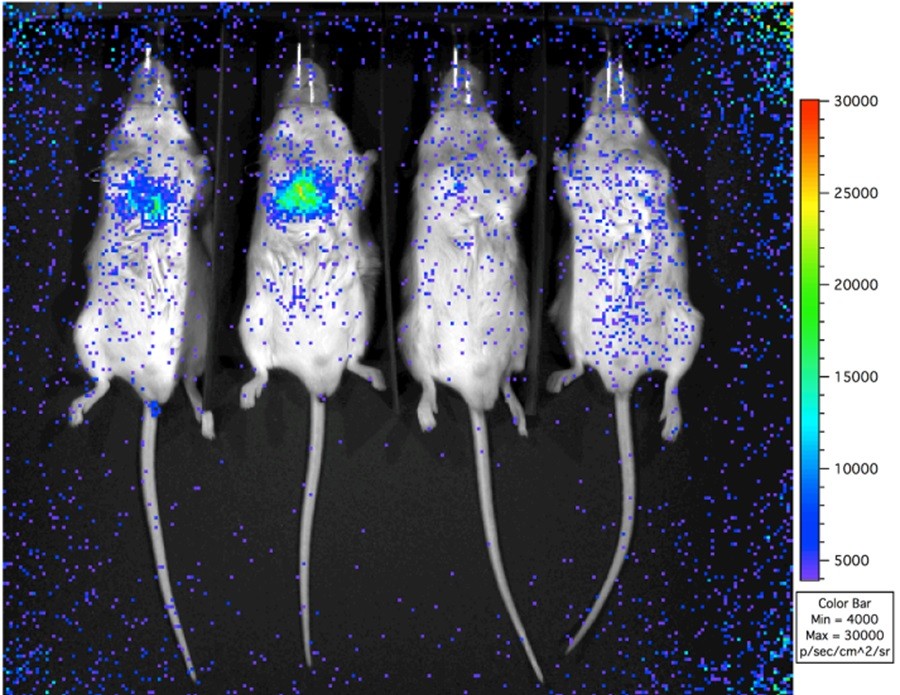
High sensitivity BLI image taken with an IVIS 100 system. 1 x 10^5 22Rv1.lucPN2 prostate cancer cells were injected intracardiacally into severe combined immunodeficient (SCID) mice. The two mice on the left had an unsuccessful IC injection whereas the two on the right were successful. A high background ‘halo’ effect is seen as a circular pattern of noisy, high-level regions where there is no real signal.
To understand the ‘halo’ effect and how it affects quantisation and detection thresholds, similar ultra-weak images were studied using the IVIS Spectrum and Spectral Instruments Imaging’s Ami HTX. For the region of the image away from the ‘halo’ effect, all three imaging systems’ lower limits were similar and capable of quantitating ultra-weak BLI signals. Near the corners of all three systems, the signal/noise for a low-level signal was lower in the IVIS 100, making it more difficult to detect a low-level signal. For the other systems, this effect was negligible, and they were able to detect ultra-weak signals across the entire field of view.
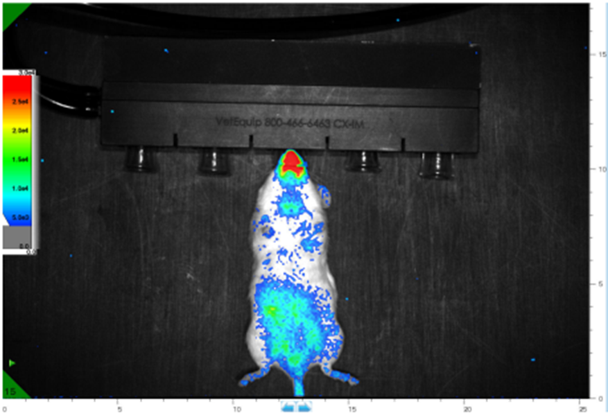
High sensitivity BLI image taken with an Ami HTX system. Photon sensitivity range is set identically to the IVIS 100 system. From the manufacturer’s specifications, the system is as sensitive as the IVIS 100 but does not exhibit the ‘halo’ effect.
The study concluded that all three instruments achieved the colour range required for high sensitivity BLI imaging, but the IVIS 200 and Ami HTX required less correction, resulting in no ‘halo’ effect.
Find out more
You can read the full study by Michael D. Henry et al. here or learn more about the Ami HTX by clicking the button below to speak to a product specialist directly.