Optimisation and evaluation of digital mammography systems.
In mammography it is essential that objects with very small contrast and diameter can be distinguished. Therefore the quality of the technical aspects of the mammography equipment should be monitored at regular time-intervals. The CDMAM 3.4 phantom is specially developed to facilitate this task in analog and digital mammography systems, i.e. detecting very low contrast and detecting very small details. The CDMAM is considered to be the world gold standard and is described in the European Protocol for quality assurance in Digital Mammography.
Measurements
To make an X-ray image, the CDMAM phantom (in combination with one or more attenuation plates) should be positioned on the bucky with the smallest disc diameters at the thorax side. Four attenuation plates with a thickness accuracy of ±1% to minimize the difference in attenuation are part of the standard delivery of the CDMAM 4.0 phantom. The images can be analysed in CDMAM 4.0 Analyser software to determine the Contrast-Detail curve and used for evaluation of the image quality.
Applications
- Quality assurance in mammography according to the 4th edition with supplements of the European Guidelines for Quality Assurance in Breast Cancer Screening and Diagnosis (3)
- For full-field digital units
- For automatic readout (human readout is possible)
- Determination if mammographic images are indicating objects with very low contrast and very small diameter
- Monitors the image information content with the Contrast-Detail curve and the spatial resolution
- Determination of the optimum exposure technique, e.g., by variation of tube potential
- Comparison of image quality at various object thicknesses by variation of the amount of PMMA
- Determination of the optimum background density
- Comparison of different mammography systems
- Study small (0.08 mm) and large (1-2 mm) diameter contrasts
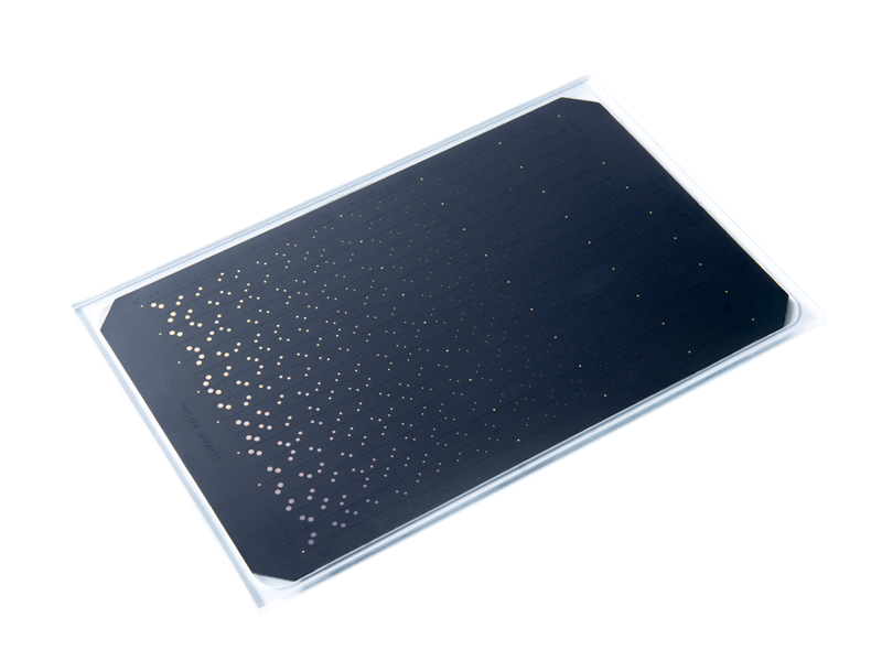
The Artinis CDMAM 4.0 phantom consists of an aluminum base with gold discs (pure gold) of various thickness and diameter. The gold discs are arranged in a matrix of 16 rows by 21 columns. Within a column the disc diameter is constant with logarithmic increasing thicknesses. The thickness is from 0.012 µm to 0.1 µm for the 2.0 mm gold discs and from 0.59 µm to 2.8 µm for the 0.08 mm gold discs. For a detailed overview of the gold disc thickness, please download our leaflet. Each square contains two identical discs (same thickness, same diameter), one in the center and one in a randomly chosen corner for human readout. The matrix grid is silkscreen printed with X-ray contrasting paint. The aluminum base (0.05 mm thick Al 1050, 99.5% pure aluminum) is attached to a cover (3 mm, PMMA). Under normal mammography-radiation conditions (Mo anode, 30 mm Mo filter, 28 kV) the aluminum base and PMMA cover together have an equivalent PMMA thickness of 10 mm. The actual attenuation of the CDMAM Phantom depends on the used PMMA thicknesses along with the phantom. The effective energy of the phantom plane will be higher when more PMMA is added to the top and bottom of the phantom.
Documents
-
Radiology-Quality-Assurance-EU-Version-1.3
Download (23.19 MB)
Catalogues
-
Radiology-Quality-Assurance-EU-Version-1.5
Download (18.37 MB)