Small animal scintigraphy using the MiE Equine Scanner
4th October 2023
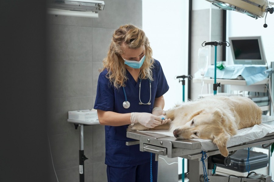
Oakham Veterinary Hospital has been using the gamma camera to nuclear image dogs referred by orthopaedic specialists
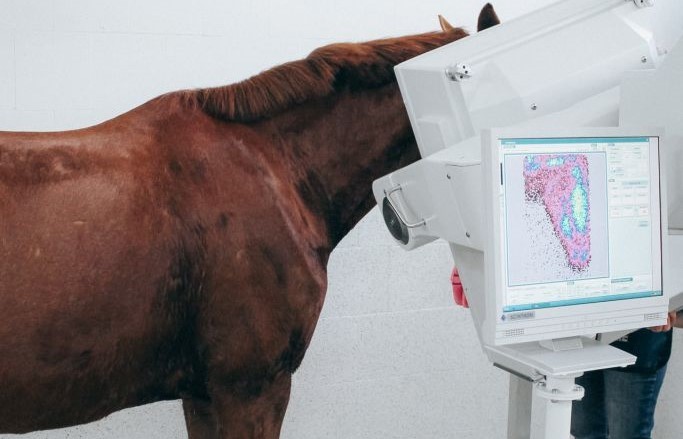
Although originally intended for equine scintigraphy, the MiE gamma camera has the necessary imaging resolution and motion correction to successfully image small animals, which is becoming increasingly common. It is being used by a global user base for renograms and p-shunt scintigraphy on dogs and in thyroid diagnostics and radioiodine treatment in cats.
Here in the UK, Oakham Veterinary Hospital has been using an MiE to nuclear image dogs diagnosed with lameness. Oakham is a Tier 3 veterinary hospital and part of the IVC Group. It offers a full suite of imaging services for both large and small animals, including scintigraphy, MRI, CT, X-ray, and Ultrasound. Being so confident in the images produced with horses, the hospital began using the MiE for dogs who were referred to Oakham by orthopaedic specialists.
“We’ve found the MiE’s scintigraphy imaging has worked particularly well on small animals.”
Ben Anghileri is the hospital’s Clinical Director. Speaking about the use of the MiE camera for small animal scintigraphy, Ben said, “The dogs were referrals from other practices that didn’t have the ability to scan. They had undiagnosed lameness with questions over their elbows and carpi. The referring orthopaedic specialist was experienced but couldn’t identify where the lameness was coming from through a clinical exam alone.
“The MiE’s scintigraphy imaging has worked particularly well on small animals, giving us functional detail about the bones and allowing us to highlight areas of bone activity. We’ve used it in conjunction with CT to give us density information about soft tissue damage.”
Shorter acquisition times with rigid registration software
MiE’s rigid registration software speeds up image acquisition, reduces the likelihood of repeating images, and eliminates the need to post-process captured images. By reducing imaging time, this reduces staff radiation dosage per examination.
Like many vets, Oakham originally used an older medical gamma camera intended for imaging humans. Ben said, “The MiE camera replaced an older system. Although it was still producing adequate images, it was coming to the end of its life, becoming increasingly unreliable and sourcing parts for servicing became difficult, so we decided to upgrade.”
The MiE uses a Nal(TI) crystal and 66 PMTs to deliver the largest field of view currently available for animal scintigraphy. Ben said, “The size of image we can acquire is much larger. I’m not confident that the old system would have coped so it’s great to be able to offer this service.”
An existing global user base
Other hospitals around the world using MiE gamma cameras for small animal scintigraphy include:
- VCA Speciality Animal Hospital in Los Angeles, USA.
- Tierklinik Norderstedt, a private clinic near Hamburg performing shunt, skeletal, renal, thyroid, and tumour scintigraphy, radioiodine therapy, and radiosynoviorthesis.
- University of Giessen near Frankfurt with two MiE cameras used exclusively in the small animal department.
- University of Vienna with two MiE cameras; one for large animal imaging and one for small.
- University of Uppsala in Sweden.
Find out more
You can learn more about the MiE equine gamma camera and small animal scintigraphy by clicking the button below to speak to a product specialist.