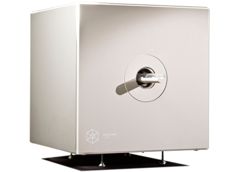
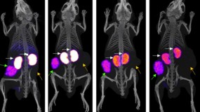
The γ-CUBE is a high-performance preclinical SPECT imager to track and quantify molecular processes

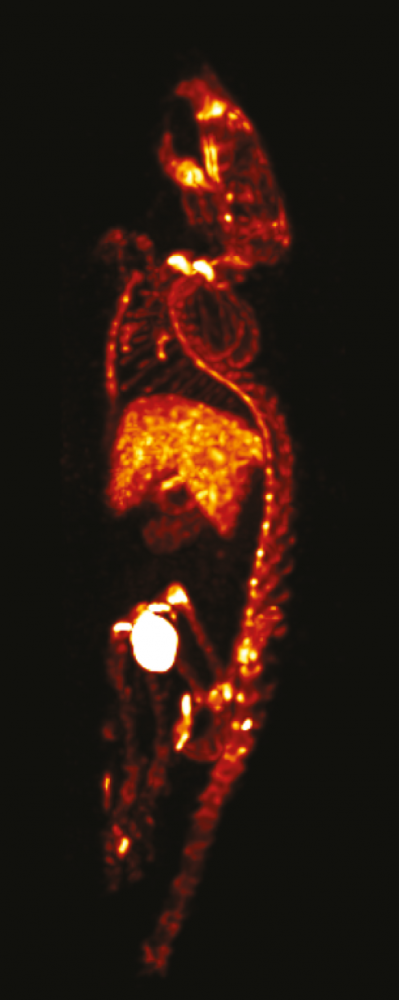
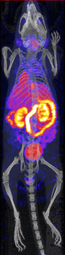
By labelling peptides, proteins, antibodies and many small molecules, with a radioisotope, virtually any molecular process can be imaged non-invasively and longitudinally with a SPECT imager.

SPECT compatible radioisotopes are typically useful for imaging longer biological half-life processes. Image and quantify the bio-distribution of monoclonal antibodies (mAbs) and peptides, (stem) cell imaging, and more.

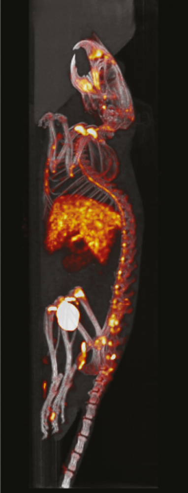
Intuitive and wireless acquisition software combined with our multimodal small animal bed allow for easy and modular multimodal imaging along with the X-CUBE (CT) and β -CUBE (PET).

γ-CUBE preclinical SPECT imager testimonials


γ-CUBE

X-CUBE

β-CUBE

  GP mouse protocol    
Field Of View
axial x transaxial
12mm x 30mm 35mm x 63mm 130mm x 72mm
Spatial Resolution
*general purpose mouse collimator
< 0,6mm 0,05mm < 1mm
Sensitivity 0,12% - > 10%
Reconstruction Code
on board GPU-based
3D MLEM, 3D OSEM FDK, ISRA FBP, 3D MLEM, 3D OSEM
Weight
over footprint of 54cm x 54cm
< 80kg < 100kg < 90kg

Existing γ-CUBE customers 

Click on the tabs below to find out more about our customers, and how they are using the γ-CUBE in their facilities. 

INRASTES, Greece

Monitoring the biodistribution of radiopharmaceuticals and gelofucine

In a study conducted by Kaloudi et al. at INRASTES, the efficacy of gelofucine in concentrating a radiopharmaceutical within tumour cells was explored, aiming to minimise damage to healthy tissues. A CCK2R antagonist, DGA1, was specifically designed and radiolabelled with 99mTc. To mitigate radiotracer accumulation in the kidneys and thus preserve healthy tissue, gelofucine was concurrently administered alongside DGA1. Using the γ-CUBE for SPECT imaging, the biodistribution of DGA1 was analysed, revealing that gelofucine successfully concentrated 99mTc in tumour tissue while reducing levels in the kidneys. This outcome effectively achieved the objective of sparing healthy tissues.

You can learn more about the study and how the MOLECUBE was used by clicking the button below.

READ MORE

Kyoto Pharmaceutical University, Japan

Studying the biodistribution of Eph receptors for cancer diagnosis

A study published by Furukawa et al. at Kyoto Pharmaceutical University has used MOLECUBES’ X-CUBE and γ-CUBE to study the biodistribution of Eph receptors, which are thought to be associated with cancer cell migration and proliferation. The benchtop preclinical imagers were used to evaluate and visualise a EphA2 receptor-specific SPECT imaging tracer for diagnosing cancer called [I-125]ETB.

You can learn more about the study and how the MOLECUBES were used by clicking the button below.

READ MORE

Hull PET Research Centre, UK

Steve Archibald: Why Hull chose MOLECUBES’ preclinical imagers for translational studies

MOLECUBES preclinical imagers have been installed at the PET Research Center (PETRC) at the University of Hull in the UK. The state-of-the-art facility uses medical imaging for research and the early diagnosis of diseases such as cancer and dementia. It supports a wide range of internationally-recognised research programmes and is part of a group of integrated clinical facilities with links to Castle Hill Hospital.

PETRC’s lab is equipped with a full suite of MOLECUBES including a γ-CUBE for SPECT, a β-CUBE for PET, and a X-CUBE for CT. By combining all three CUBES, the limitation of each modality is offset and provides a multimodal approach to preclinical imaging.

Watch our video with Professor of Molecular Imaging Steve Archibald or click the button below to find out more.

READ MORE

BIOEMTECH, Greece

BIOEMTECH, Greece

Prof. George Loudos is the founder and CEO of BIOEMTECH. BIOEMTECH labs, are located in Athens and support the complete chain of pre-clinical studies for testing new  compounds  including: animal hosting premises (mice and rats), in vitro lab for cell studies, radiochemistry lab for labelling various compounds and multimodal imaging facility,  including fast  dynamic molecular screening, 3D SPECT/CT systems (x/γ CUBE, Molecubes), plus a PET and optical system, developed in house. 

Why Choose molecubes?

BIOEMTECH has an academic origin and deep technical background, since we have launched the first in vivo molecular screening systems (the eyes) for whole body planar dynamic imaging. We chose Molecubes because of the teams’ academic background and thus deep knowledge of the technology and applications. Knowing the core team and their progress for more than a decade, we felt from the first moment absolute trust in their proposed solutions but also in their support and innovative ideas.

How would you rate your experience with Molecubes?

Having two of the Molecubes systems for almost 2 years now, we have deeply appreciated the end user experience and the continuous support.

What specific feature do you like the most?

Some of the features we have mostly appreciated is the wide range of applications that can be covered, along with a great simplicity of use, beautiful images and accessibility and promptness of the supporting team.

Which applications do you run on the system?

Up to now, we have used the systems for multiple SPECT applications on cardiac imaging, several oncology applications, lung imaging, brain targeting and a wide range of nanoparticle studies. On CT, besides animals, we have imaged multiple stained tissues with great accuracy, as well as dental tissues and in vivo bone studies.

Would you choose for benchtop instruments again?

It is in our plans to also purchase the β-CUBE and thus have the complete CUBES platform in our pre-clinical labs.

How often are the instruments used?

The instruments are used on a weekly basis and during busy periods daily. Acquisitions and reconstructions are running in parallel for a higher throughput. Since our throughput is high, we look forward to seeing new  improvements  related to multiple animal imaging or remote workstations for parallel post-processing of the acquired data, greatly saving experimental time.

Indiana University School of Medicine/IUPUI, USA

Indiana University School of Medicine

"To achieve the goals outlined in the MODEL-AD and TREAT-AD grants, the Territo lab will be scanning between 800-1400 mice per year. The team at IU was looking for a system, which has high sensitivity, resolution, and uniformity along with well-established and easy to follow workflows that would facilitate rapid multi-animal acquisition and reconstructions."

Prof. Paul Territo, Indiana University School of Medicine

NYU Grossman School of Medicine, USA

Imaging in a BSL-3 lab for infectious diseases

The Preclinical Imaging Laboratory at the NYU Grossman School of Medicine, as part of the Division for Advanced Research Technologies within the Office of Science & Research, enables NYU biomedical scientists to image live animals on an organ, tissue, cell, or molecular level using state-of-the-art preclinical imaging technologies. The lab is equipped with MOLECUBES benchtop preclinical imagers including a γ-CUBE for SPECT, a β-CUBE for PET, and two X-CUBEs for CT, one of which was originally procured specifically for infectious diseases research including COVID-19.

We spoke to Director of the Preclinical Imaging Laboratory and Associate Professor Youssef Wadghiri about his work using the CUBES. You can find out what he had to say by clicking the button below. 

READ MORE

Documents

The LabLogic Group will always treat your details with the utmost care and will never sell them to third party companies.

Please choose one of the following:


Previous
Next